


What is MRI?
Magnetic resonance imaging (MRI), nuclear magnetic resonance imaging (NMRI), or magnetic resonance tomography (MRT) is a medical imaging technique used in radiology to visualize internal structures of the body in detail. MRI makes use of the property of nuclear magnetic resonance (NMR) to image nuclei of atoms inside the body.
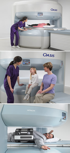
An MRI scanner is a device in which the patient lies within a large, powerful magnet where the magnetic field is used to align the magnetization of some atomic nuclei in the body, and radio frequency magnetic fields are applied to systematically alter the alignment of this magnetization. This causes the nuclei to produce a rotating magnetic field detectable by the scanner and this information is recorded to construct an image of the scanned area of the body. Magnetic field gradients cause nuclei at different locations to precess at different speeds, which allows spatial information to be recovered using Fourier analysis of the measured signal. By using gradients in different directions 2D images or 3D volumes can be obtained in any arbitrary orientation.
MRI provides good contrast between the different soft tissues of the body, which makes it especially useful in imaging the brain, muscles, the heart, and cancers compared with other medical imaging techniques such as computed tomography (CT) or X-rays. Unlike CT scans or traditional X-rays, MRI does not use ionizing radiation.
The use of X-rays, a type of ionizing radiation, by computed tomography (CT) scanner, to acquire images, make computed tomography a good tool for examining tissue composed of elements of a higher atomic number than the tissue surrounding them, such as bone and calcifications (calcium based) within the body (carbon based flesh), or of structures (vessels, bowel). MRI, on the other hand, uses non-ionizing radio frequency (RF) signals to acquire its images and is best suited for soft tissue (although MRI can also be used to acquire images of bones, teeth and even fossils).
CT scans use ionizing radiation (X-rays) to produce images, which can damage DNA and subsequently cause cancer. There is a small increased risk of cancer with CT scans. It is estimated that 0.4% of current cancers in the United States are due to CTs performed in the past and that this may increase to as high as 1.5-2% with 2007 rates of CT usage. Unlike CT, MRI does not use ionizing radiation, though it is associated with other risks.
Contrast in CT images is generated purely by X-ray attenuation, while a variety of properties may be used to generate contrast in MR images. By variation of scanning parameters, tissue contrast can be altered to enhance different features in an image (see Applications for more details). Both CT and MR images may be enhanced by the use of contrast agents. Contrast agents for CT contain elements of a high atomic number, relative to tissue, such as iodine or barium, while contrast agents for MRI have paramagnetic properties, such as gadolinium and manganese, used to alter tissue relaxation times. Commonly used MRI contrast agents may be contraindicated in persons with significant permanent or transient kidney dysfunction.
CT and MRI scanners are able to generate multiple two-dimensional cross-sections (tomographs, or "slices") of tissue and three-dimensional reconstructions. MRI can generate cross-sectional images in any plane (including oblique planes). In the past, CT was limited to acquiring images in the axial plane (or near axial). The scans used to be called Computed Axial Tomography scans (CAT scans). However, the development of multi-detector CT scanners with near-isotropic resolution, allows the CT scanner to produce data that can be retrospectively reconstructed in any plane with minimal loss of image quality. For purposes of tumor detection and identification in the brain, MRI is generally superior. However, in the case of solid tumors of the abdomen and chest, CT is often preferred as it suffers less from motion artifacts. Furthermore, CT usually is more widely available, faster, and less expensive. However, CT has the disadvantage of exposing the patient to harmful ionizing radiation.
MRI is also best suited for cases when a patient is to undergo the exam several times successively in the short term, because, unlike CT, it does not expose the patient to the hazards of ionizing radiation. However MRI is usually contraindicated if the patient has any type of medical implant, such as vagus nerve stimulators, implantable cardioverter-defibrillators, loop recorders, insulin pumps, cochlear implants, deep brain stimulators, etc.; metallic foreign bodies such as shrapnel or shell fragments; or metallic implants such as surgical prostheses. These devices can malfunction or heat up during a scan, and as such, for patients having them, CT scans are considered the safer option.
Patient Resources